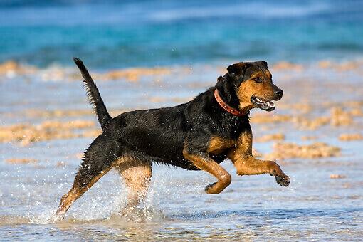
As a puppy, Champ snuck into Mary’s knitting bag and swallowed a small ball of yarn. When he became unwell, Mary called our practice right away. The yarn didn’t show up on the x-ray so we used endoscopy to look inside Champ’s digestive tract and remove the yarn. Endoscopy is a procedure that uses a tiny video camera at the end of a very narrow scope to see the inside of the body. Sometimes our practice uses endoscopy for surgical procedures but most often we use it for diagnostic purposes. Minimally invasive, the procedure requires relatively little recovery.
Before the endoscopy, our practice will first perform a full physical exam. Because of the use of anesthesia, your pet may require blood tests to ensure sure the ability to properly metabolize the medications. Our practice then administers the anesthesia and carefully monitors your pet during the entire procedure. After the endoscopy, we gently awaken your pet from the anesthesia and in most cases, you can take your pet home the same day.
Sometimes our practice uses endoscopy to perform biopsies, take cultures, or remove tumors, masses, or foreign bodies like Champ’s ball of yarn. After the procedure, our practice evaluates the information gathered or possibly runs lab tests on the tissue retrieved.
Endoscopy allows us to view many different parts of the body. For example, if you notice your pet experiencing respiratory issues, we may use endoscopy to look inside of their nose and sinuses to determine if an infection is present, the possibility of a tumor or if your pet inhaled a foreign body. In Champ’s case, we used endoscopy to travel through the intestines and find the yarn.
Some common areas of endoscopy include:
- Gastrointestinal endoscopy- Our practice uses a very long and flexible scope. We direct the scope to travel down the intestines which enables biopsy, the removal of foreign bodies, or to diagnosis of GI issues such as polyps or colitis.
- Bronchoscopy- Allows our practice to view the throat and lungs and identify issues such as polyps, foreign bodies, or lung cancer.
- Rhinoscopy- We use a flexible scope to view the sinuses, and remove foreign bodies, inflammation, or fungal infections. Anterior rhinoscopy advances a flexible scope through the nose, posterior advances the scope through the mouth to examine the back of the nasal cavity.
- Cystoscopy- Our practice can view the urinary tract, including the urethra and bladder. We can identify and sometimes remove kidney and bladder stones.
